Bones In Leg Diagram : Leg Fracture What You Need To Know. The femur is the single bone of the thigh. 2006 kia optima belt diagram. The bones of the leg are the femur, tibia, fibula and patella.the foot bones shown in this diagram are the talus, navicular, cuneiform, cuboid, metatarsals and calcaneus. Medical anatomy human skeletal system anatomy bones radiology student human body anatomy medical knowledge nursing students physiology biology. The major bones of the leg are the femur (thigh bone), tibia (shin bone), and adjacent fibula, and these are all long bones.
Learn vocabulary, terms, and more with flashcards, games, and other study tools. The tibia and fibula are two long bones that run parallel to each other, forming the scaffold of the leg and providing attachment points for many muscles. 2006 kia optima belt diagram. He leg's main function in the human is for locomotion and support of the rest of the body. Inside of a female human body bones 12 photos of the inside of a female human body bones , bone.
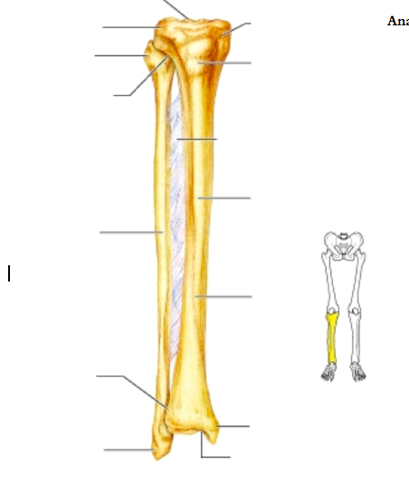
Leg Anatomy from fpnotebook.com The tibia and fibula form the ankle joint with the talus,. Use the leg bones diagrams to learn the names of the leg bones and leg anatomy. The bones of the leg and foot form part of the appendicular skeleton that supports the many muscles of the lower limbs. The tibia is much larger than the fibula and bears almost all of the body's weight. This area is commonly referred to as the calf. Bone diagram forehead (frontal bone) nose bones (nasals) cheek bone (zygoma) upper jaw (maxilla) lower jaw (mandible) breast bone (sternum) upper arm bone (humerus) lower arm bone (ulna) thigh bone (femur) collar bone (clavicle) toe bones (phalanges) ankle bones (tarsals) kneecap (patella) shin bone The bones of the leg are the femur, tibia, fibula and patella. Human leg bones, computer artwork.
The bones of the leg are the femur, tibia, fibula and patella.the foot bones shown in this diagram are the talus, navicular, cuneiform, cuboid, metatarsals and calcaneus.
Most of the leg skeleton has bony prominences and margins that can be palpated and some serve as anatomical landmarks that define the extent of the leg. Its lower end helps create the knee joint. At the same time, the bones and joints of the leg and foot must be strong enough to support the body's weight while remaining. Bone diagram forehead (frontal bone) nose bones (nasals) cheek bone (zygoma) upper jaw (maxilla) lower jaw (mandible) breast bone (sternum) upper arm bone (humerus) lower arm bone (ulna) thigh bone (femur) collar bone (clavicle) toe bones (phalanges) ankle bones (tarsals) kneecap (patella) shin bone Use the leg bones diagrams to learn the names of the leg bones and leg anatomy. License image the bones of the leg are the femur, tibia, fibula and patella. The patella (kneecap) is the sesamoid bone in front of the knee. Home » unlabelled » bones in leg diagram / your leg bones are very large and strong to help support the weight of your body. The knee joint is the largest joint in the body and is primarily a hinge joint, although some sliding and rotation occur. 2006 kia optima belt diagram. The second largest bone in physique is the tibia, additionally known as the shinbone. The patella is the kneecap and articulates with the distal femur. The hip itself is a ball and socket joint, much like the shoulder.the structures necessary to create this joint are the socket, the joint capsule, muscle, ligaments, and the neck.
Inside of a female human body bones. The human leg consists of 8 bones, 4 per leg. Stephen longenecker, an orthopedic surgeon at the reading hospital and medical center, decribes 23 april 2001 in reading how an external fixation. The pubis, ischium, and ilium together constitute the pelvis while the thigh bone is the femur. The fibula is mainly a muscle attachment point and is used to help maintain balance.
Lower Leg Bones Diagram Quizlet from o.quizlet.com The pubis, ischium, and ilium together constitute the pelvis while the thigh bone is the femur. The movements your muscles make are coordinated and controlled by the brain. Disposition of rotator cuff muscles diagram. The human leg consists of 8 bones, 4 per leg. The tibia, commonly known as the 'shin bone', is the largest and most medial of the two.you can palpate its anterior border when you run your finger down the anterior aspect of your leg. The lower limb contains 30 bones. Leg bones, learn what and where these are as well as their functions and how we use them. Learn vocabulary, terms, and more with flashcards, games, and other study tools.
At the distal end of the femur, two rounded condyles meet the tibia and fibula bones of the lower leg to form the knee joint.
Joints of hand anterior view, lateral view, right hand. An easy source to get a ton of information easily. Leg bones, learn what and where these are as well as their functions and how we use them. The patella is the kneecap and articulates with the distal femur. Bones pain hand and arm bones diagram. Learn vocabulary, terms, and more with flashcards, games, and other study tools. The thigh bone, or femur, is the large upper leg bone that connects the lower leg bones (knee joint) to the pelvic bone (hip joint). Most of the leg skeleton has bony prominences and margins that can be palpated and some serve as anatomical landmarks that define the extent of the leg. The lower leg extends from the knee to the ankle. Its lower end helps create the knee joint. The knee joint is the largest joint in the body and is primarily a hinge joint, although some sliding and rotation occur. The femur is the single bone of the thigh. Bones pain hand and arm bones diagram.
These bones have a marrow, but not a bone marrow cavity. The tibia and fibula are the bones of the lower leg. The thigh bone, or femur, is the large upper leg bone that connects the lower leg bones (knee joint) to the pelvic bone (hip joint). Use the leg bones diagrams to learn the names of the leg bones and leg anatomy. These are the femur, patella, tibia, fibula, tarsal bones, metatarsal bones, and phalanges (see figure 6.51).
Leg Picture Image On Medicinenet Com from images.medicinenet.com The tarsal bones in the foot are located amongst tibia, metatarsal bones, and fibula. These are the femur, patella, tibia, fibula, tarsal bones, metatarsal bones, and phalanges (see figure 6.51). The lower limb contains 30 bones. The diagram of bones in the ankle and foot is given below: Stephen longenecker, an orthopedic surgeon at the reading hospital and medical center, decribes 23 april 2001 in reading how an external fixation. He leg's main function in the human is for use the leg bones diagrams to learn the names of the leg bones and leg anatomy. There are in all 7 bones, which fall under tarsal bones category. Bone diagram forehead (frontal bone) nose bones (nasals) cheek bone (zygoma) upper jaw (maxilla) lower jaw (mandible) breast bone (sternum) upper arm bone (humerus) lower arm bone (ulna) thigh bone (femur) collar bone (clavicle) toe bones (phalanges) ankle bones (tarsals) kneecap (patella) shin bone
Its lower end helps create the knee joint.
Bone structure horse hind leg 12 photos of the bone structure horse hind leg , bone. The bones of the hip include the femur, the ilium, the ischium, and the pubis. (note, the radius and ulna bones also have this membrane.) this membrane keeps the tibia and fibula together and provides strength and stability for them. Diagram and names of leg bones, diagram of foot and leg bones, diagram of leg bones, diagram of lower leg bones, diagram of the bones in your leg, bone, diagram and. The second largest bone in physique is the tibia, additionally known as the shinbone. He leg's main function in the human is for use the leg bones diagrams to learn the names of the leg bones and leg anatomy. The patella is the kneecap and articulates with the distal femur. The diagram of bones in the ankle and foot is given below: The bones of the leg and foot form part of the appendicular skeleton that supports the many muscles of the lower limbs. The bones of the leg are the femur, tibia, fibula and patella.the foot bones shown in this diagram are the talus, navicular, cuneiform, cuboid, metatarsals and calcaneus. Most leg pain results from wear and tear, overuse, or injuries in joints or bones or in muscles, ligaments, tendons or other soft tissues. Related posts of bones leg diagram picture bone structure horse hind leg. The tarsal bones in the foot are located amongst tibia, metatarsal bones, and fibula.
Share :
Post a Comment
for "Bones In Leg Diagram : Leg Fracture What You Need To Know"


Post a Comment for "Bones In Leg Diagram : Leg Fracture What You Need To Know"